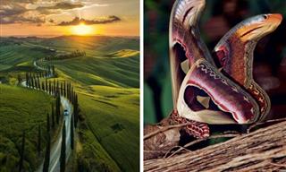
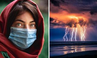
The Nikon International Small World Competition began all the way back in 1975. It was intended to recognize the efforts of those involved with photography through the light microscope. During the time since, it has become one of the world's leading showcases for photomicrography, and features images from the widest array of scientific disciplines. Here are this year's finalists and winners:
Click on images to enlarge
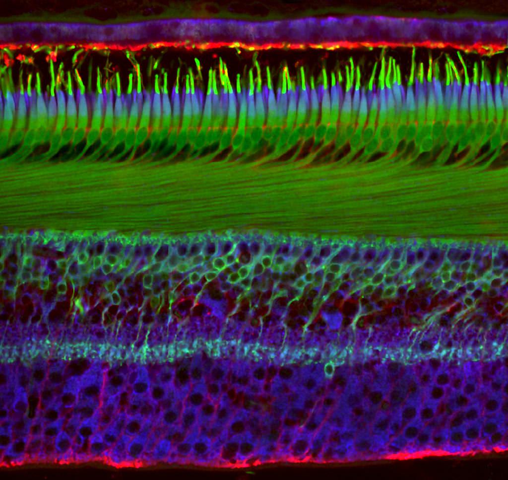
20. Human Retina - Dr. Nicolas Cuenca & Isabel Ortuno-Lizaran
- Technique
- Immunocytochemistry and Confocal Microscopy
- Magnification
- 40x (objective lens magnification)
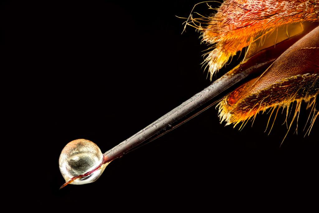
19. Asian hornet with venom on stinger - Pierre Anquet
- Technique
- Reflected Light, Focus Stacking
- Magnification
- 6.3x (objective lens magnification)
-
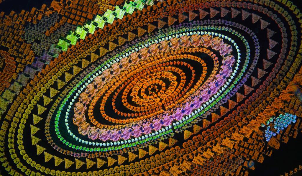
18. Amino acid crystals - Justin Zoll
- Technique
- Polarized Light, Image Tiling
- Magnification
- 4x (objective lens magnification)
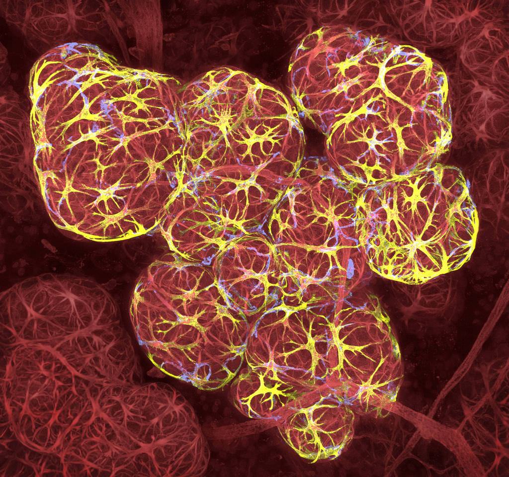
17. Breast tissue in lactation - Caleb Dawson
- Technique
- 3D Confocal Microscopy
- Magnification
- 63x (objective lens magnification)
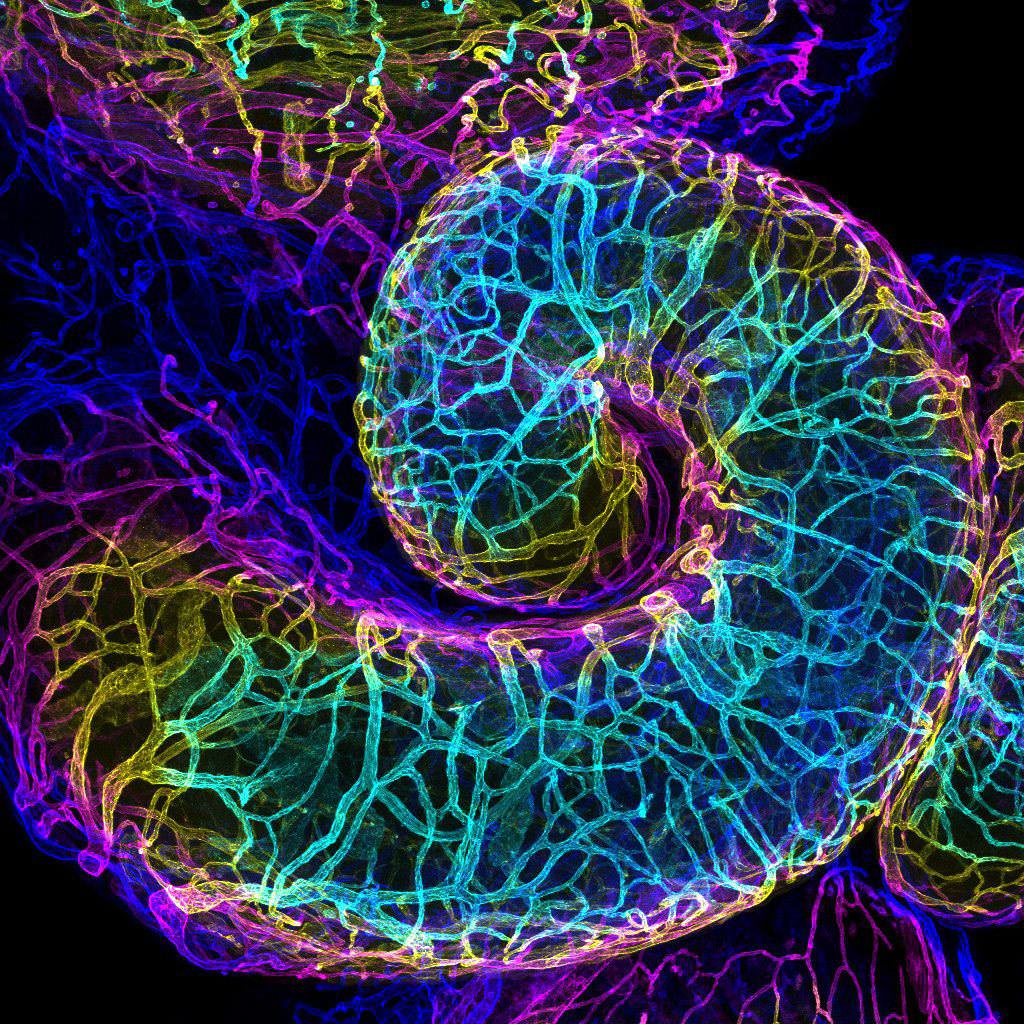
16. Mouse oviduct vasculature - Dr. Amanda D. Phillips Yzaguirre
- Technique
- Confocal
- Magnification
- 10x (objective lens magnification)
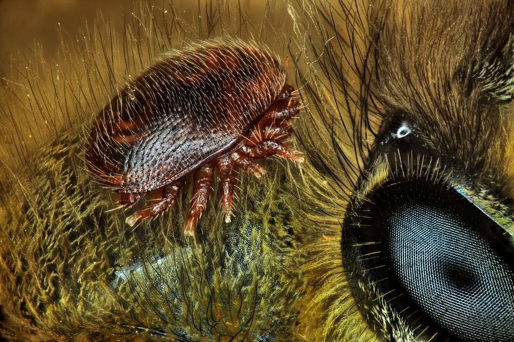
15. Mite on the back of a honeybee - Antoine Franck
- Technique
- Focus Stacking
- Magnification
- 1x (objective lens magnification)
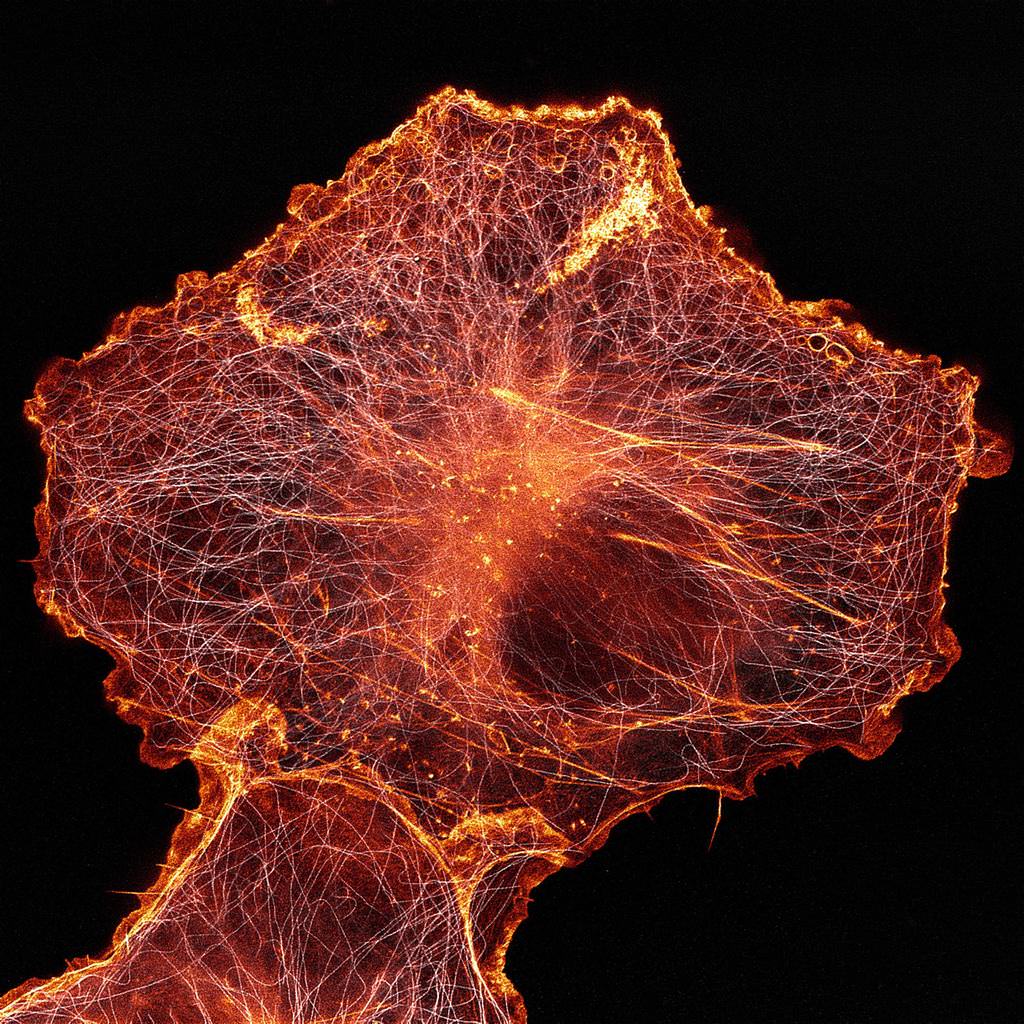
14. African green monkey cell (COS-7) stained for actin and microtubules - Andrew Moore and Dr. Erika Holzbaur
- Technique
-
Stimulated Emission Depletion (STED) Microscopy
- Magnification
- 100x (objective lens magnification)
13. Acorn barnacle - Charles B. Krebs
- Technique
- Autofluorescence
- Magnification
- 5x (objective lens magnification)
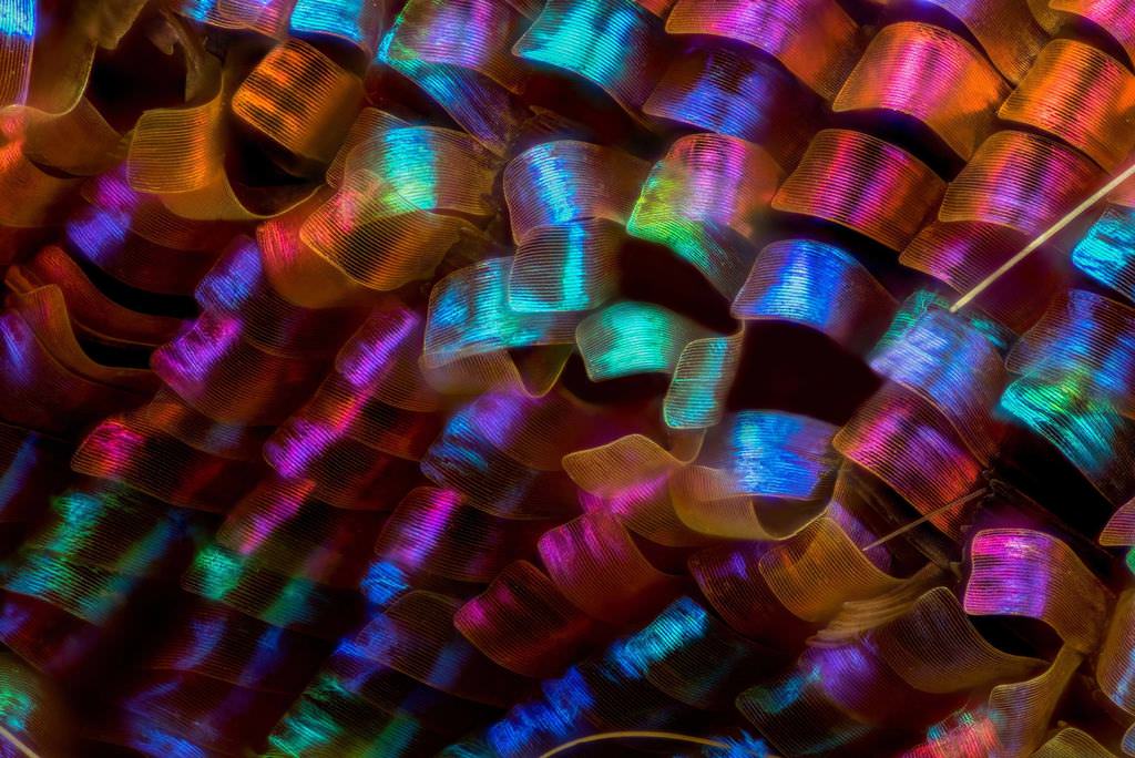
12. Butterfly wing scales - Luciano Andres Richino
- Technique
- Image Stacking
- Magnification
- 20x (objective lens magnification)
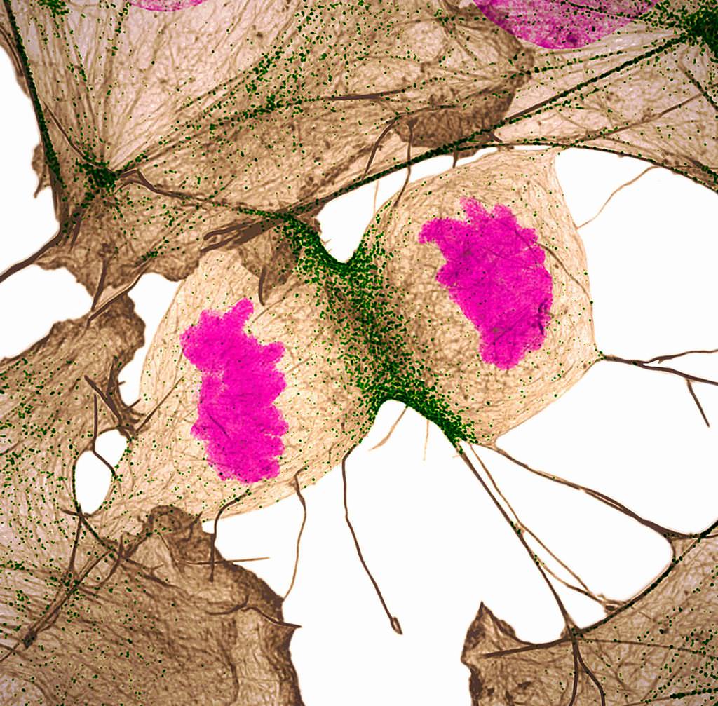
11. Human fibroblast dividing, showing actin (gray), myosin II (green) and DNA (magenta) - Nilay Taneja and Dr. Dylan Burnette
- Technique
- Structured Illumination Microscopy
- Magnification
- 60x (objective lens magnification)
10. Stalks with pollen grains - Dr. Csaba Pinter
- Technique
- Focus Stacking
- Magnification
- 3x (objective lens magnification)
9. Security hologram - Dr. Haris Antonopoulos
- Technique
- Darkfield Epi-illumination
- Magnification
- 10x (objective lens magnification)
8. Mango seed weevil - Pia Scanlon
- Technique
- Stereomicroscopy, Image Stacking
- Magnification
- 1x (objective lens magnification)
7. Human tear drop - Norm Baker
- Technique
- Darkfield
- Magnification
- 5x (objective lens magnification)
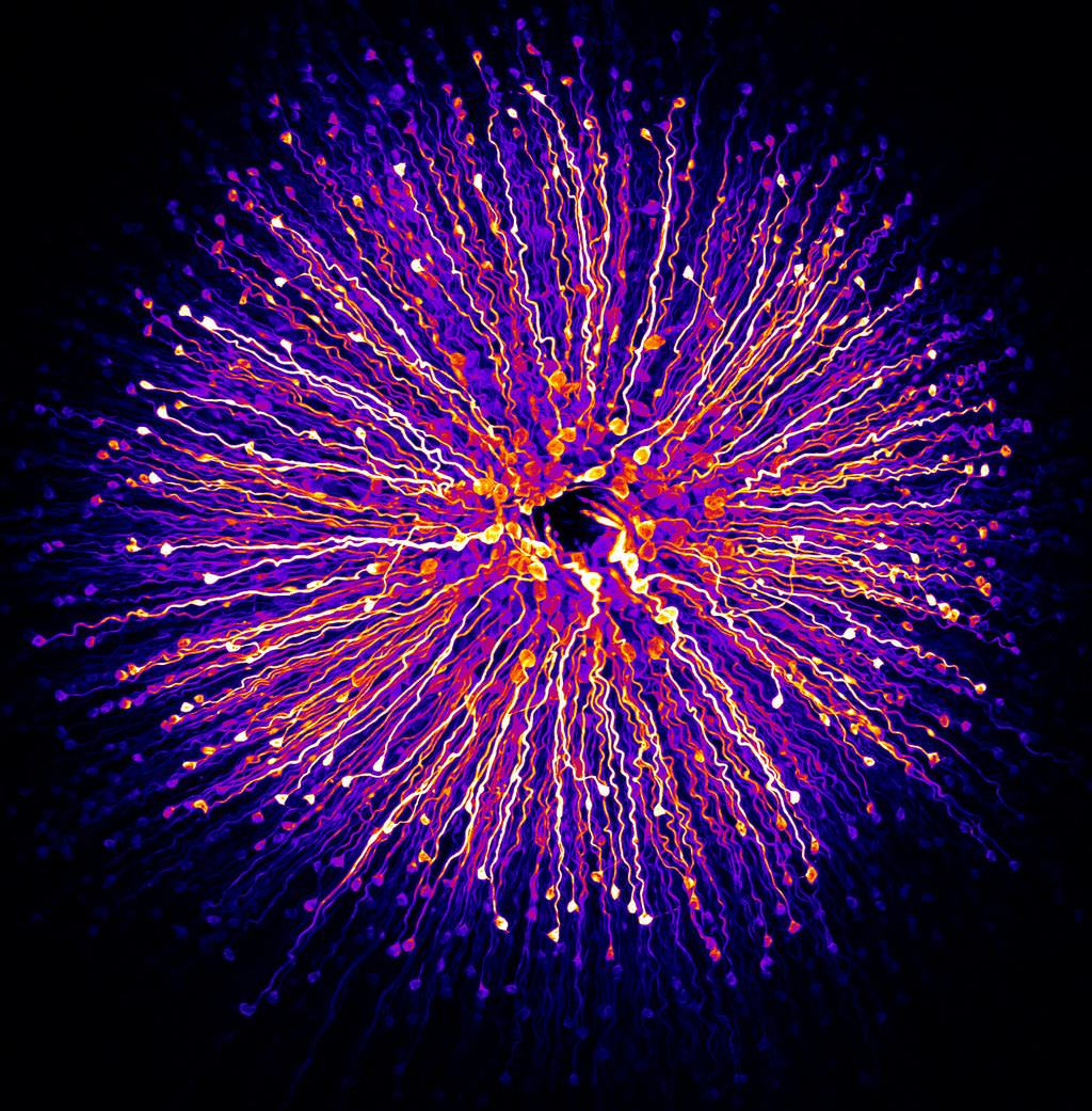
6. Central region of the retina - Hanen Khabou
- Technique
- Fluorescence
- Magnification
- 40x (objective lens magnification)
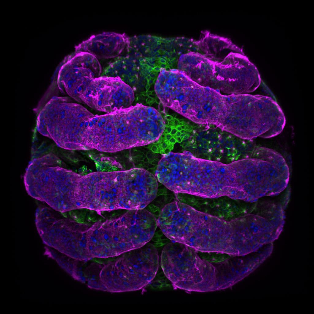
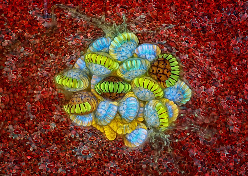
5. Spider embryo stained in different colors - Dr. Tessa Montague
- Technique
- Confocal
- Magnification
- 20x (objective lens magnification)
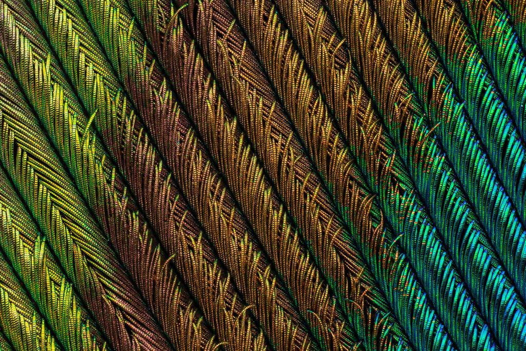
4. Peacock feather section - Can Tuncer
- Technique
- Focus Stacking
- Magnification
- 5x (objective lens magnification)
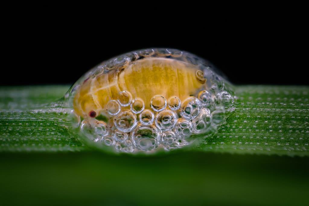
3. Spittlebug nymph in its bubble house - Saulius Gugis
- Technique
- Focus Stacking
- Magnification
- 5x (objective lens magnification)
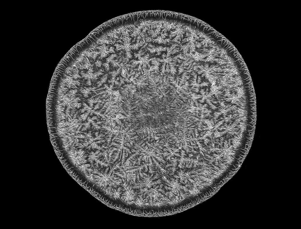
2. Fern sorus - Rogelio Moreno Gill
- Technique
- Autofluorescence
- Magnification
- 10x (objective lens magnification)
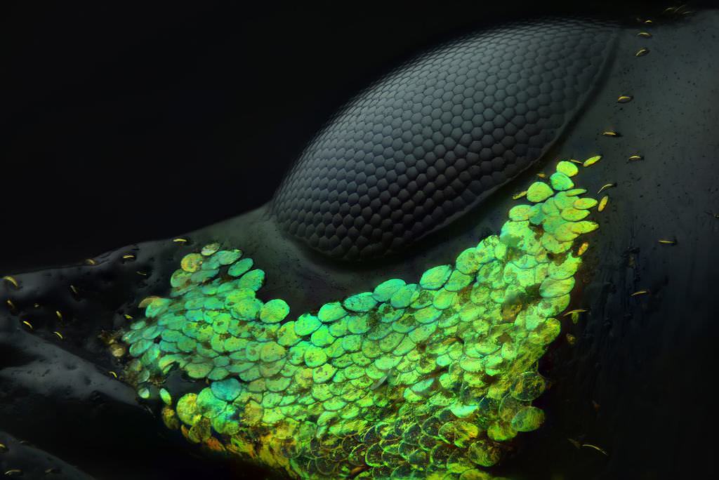
1. Eye of a Metapocyrtus subquadrulifer beetle - Yousef Al Habshi