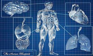
Here's a bit of news you probably weren't expecting to hear today: After more than 100 years of X-rays being flat and monochrome, they may very soon finally take the leap into the 21st Century and start being displayed as full-color 3D models.
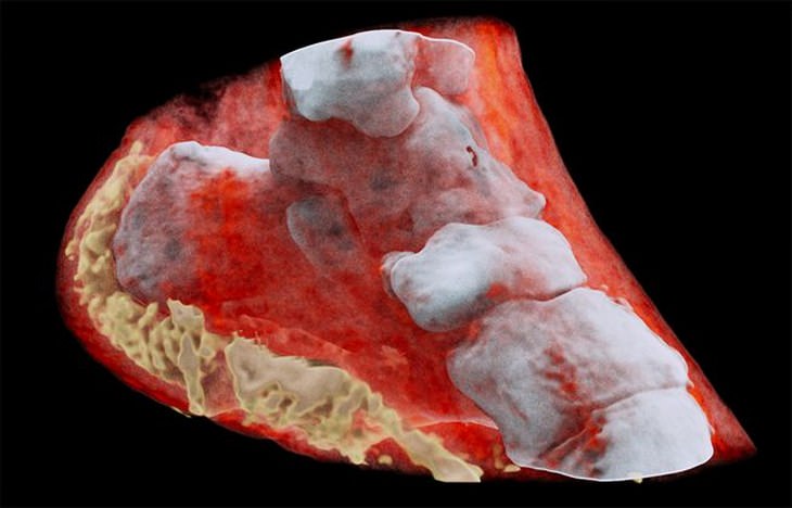
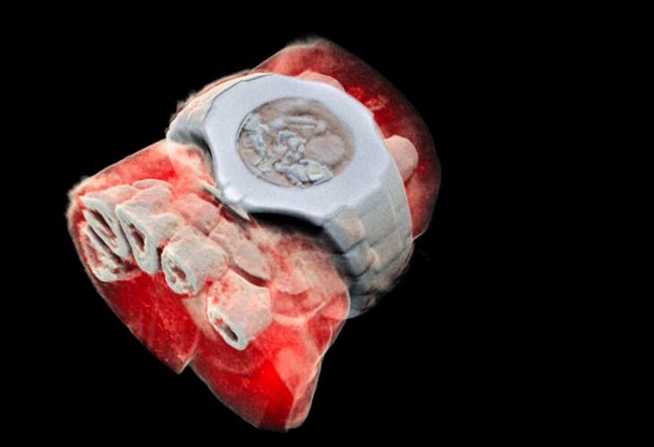
Phil and Anthony Butler, father and son scientists from the universities of New Zealand's Canterbury and Otago have created the world's first ever 3D color medical scanner. They named it the MARS spectral X-ray scanner, and it is able to capture internal images of both bone as well as all of its surrounding tissue.
According to Anthony Butler, "X-ray spectral information allows health professionals to measure the different components of body parts such as fat, water, calcium, and disease markers," and that "traditional black-and-white X-rays only allow measurement of the density and shape of an object."
As you can see from the video below, the scanner will also allow doctors to see the inner structure of a target area layer by layer:
Whereas regular X-rays use film or a sensor to record radiation levels, the MARS scanner actually measures the whole X-ray spectrum, and even goes as far as to detect and count the many particles as they collide with the pixels. This means that the MARS scanner works in a similar way to a camera, providing an HD image that is in color and fully 3D.
The technology used in the MARS scanner comes straight from CERN's Large Hadron Collider, which is the biggest and most powerful particle accelerator in the world. In fact, a chip which was designed for particle detection and imaging in the collider, known as Medipix3, was actually adapted by Phil and Anthony Butler's team so that it could be used in the MARS scanner.
According to Phil Butler, "this technology sets the machine apart diagnostically because its small pixels and accurate energy resolution mean that this new imaging tool is able to get images that no other imaging tool can achieve."
While the MARS scanner has been under development for close to ten years, clinical trials have only begun very recently. However, all results so far are showing that the MARS scanner will very likely become one of the medical community's most indispensable tools in the near future.
"So far researchers have been using a small version of the MARS scanner to study cancer, bone, and joint health, and vascular diseases that cause heart attacks and strokes," Butler said. "In all of these studies, promising early results suggest that when spectral imaging is routinely used in clinics it will enable more accurate diagnosis and personalization of treatment."
Don't forget to share this exciting news with your friends and family, too!
Source: mnn
Images: marsbioimaging
Related Articles:
Bone Spurs: What They are and How to Treat Them