|
Life is in the details, and these stunning microscopic images prove it! In the recent Nikon Small World Competition, more than 2,000 different images were submitted from 80 different countries, with all of the contenders hoping to win the glorious title of the world's best small photographer. The winner of the competition was Wim van Egmond from the Netherlands who photographed a marine diatom, a colonial plakton organism. Although his image is surely gorgeous, there were many other outstanding images presented to the judges that could have equally one the prize for the most amazing and break-taking small photos. These photos are sure to enlighten the mind and senses to the beauty of our microscopic world. |
|
A Ladybug's Foot |
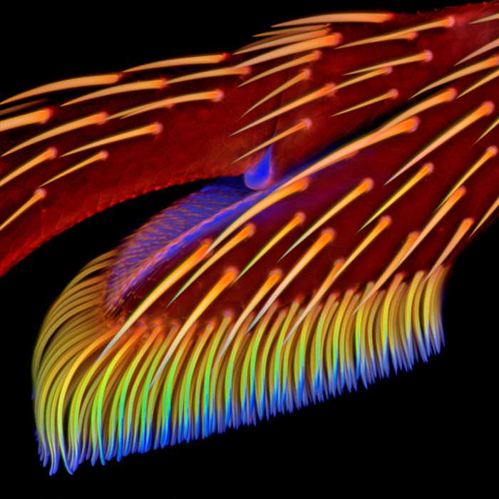 |
| This microscopic photograph of the foot of a ladybug, or a Coccinellidae in scientific language, was taken at a 20x microscopic zoom and was colored fluorescently for a beautiful effect. The ladybug is a common beatle that usually has scarlet, orange or yellow wing covers with black dots on them. The image magnifies the leg of the ladybug to between understand how these tiny creatures are able to grasp on to things so tightly. Image by: Jan Michels |
|
|
|
Moss and Bacteria |
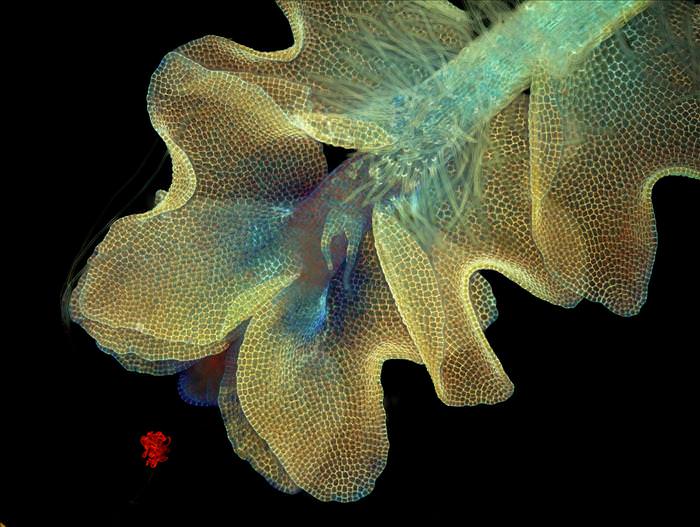 |
| An image of liverwort moss, a common green leafy moss, chasing after a cyanobacteria in the process of photosynthesis. The image was captured at a 50x magnetic zoom and used UV light to create the florescent look. Cyanobacteria is similar in its genetic structure to the chloroplasts that are responsible for making plants green, and reacts in a symbiotic manner with these other photosynthesizing agents. In short, this piece of moss is eagerly trying to get his green fix! Image by: Magdalena Turzanska |
|
|
|
The Eye of A Ghost Shrimp |
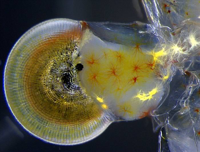 |
| Also known as the glass shrimp, these mostly transparent bottom dwellers are known for being excellent scavengers. They reach a maximum size of 2 inches, which is why a specially strong zoom of 140x was used to take a picture of this creature's eye. The photograph was also taken using a special microscope called a steromicroscope, which is able to 'dissect' the object and recreate it in a 3D magnified version. Here, you can see how the retinal nerves connect to the eye itself , and the ones that are lit are transferring those images to the shrimps brain. Image by: Vitoria Tobias |
|
|
|
A Mouse Embryo |
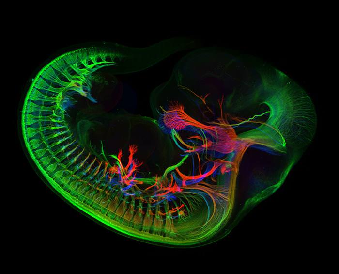 |
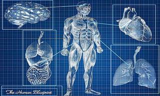
| At first glance this may look like a regular human embryo, but it is really the embryo of our far-off cousin, the mouse. Mice begin reproducing when they are only 50 days of age, and female mice produce on average 10-12 pups per cycle. This mouse has yet to be born, but after 20 days, it will leave its mother's womb with his brothers and sisters and beings its life like any other mammal. This image captures the intricacy of the mouse's young nervous system with a confocal lense that differentiates in contrast according to the density of the object it captures. Image by: Zhong Hua |
|
|
|
Mouse Vertebra Section |
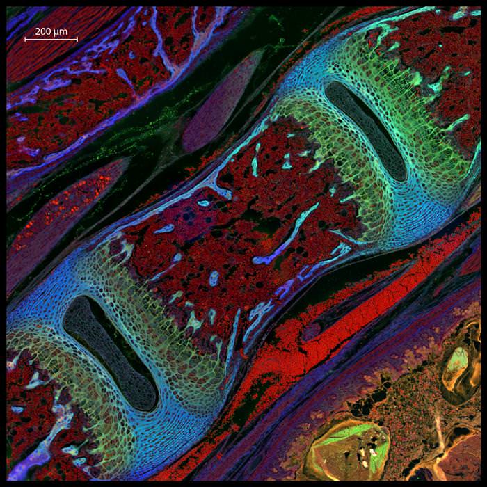 |
| Another close up image from the world of mice, this stunning photograph was taken at a magnification of 200x! The nervous system of the mouse is eerily similar to that of humans, which is one of the reasons that they are used in neurological research. Many times, scientists create what are called 'knockout' mice, which are genetically engineered with a particular neurological disease like Parkinson's or Alzheimer's, and examined for the exact changes in their nervous system. Image by: Michael Paul Nelson and Samantha Smith |
|
|
|
Sugar Transport in Fat Cells |
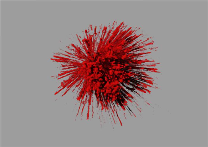 |
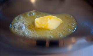
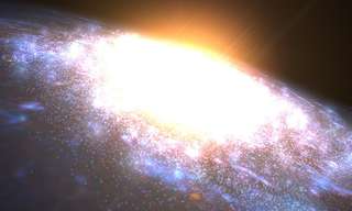
| This unique image captures the moment that sugar, or glucose, enters the fat cell. Once the glucose molecule breaks through the fat cell membrane it seems to explode in uncontrollable excitement. The image was taken at an amazing magnification of 1,000,000x inside a living fat cell that blows up (literally) with a sugar burst. Image by: James Burchfield |
|
|
|
The Retina of a Painted Turtle |
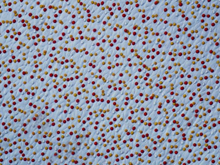 |
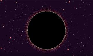
| The retina is the layer of light sensitive tissue over the eye, and in the case of this particular image, over the eye of the painted turtle. The painted turtle is native to North America and is noted for its colorful body parts like yellow bellies and red arms. This image of the turtle's retina seems to harken at its colorful nature. Image by: Joseph Corbo |
|
|
|
The Marine Diatom |
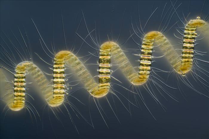 |
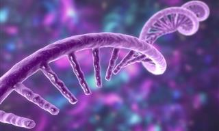
| This colonial plankton organism known scientifically as the Chaetoceros is a two-celled marine organism. Scientists have long tried to recreate its genetic pattern, which seems ironic as its shape so resembles a DNA double helix. The image one first place in the Nikon Small World Photomicrography Competition and was taken at a magnification of 250x. Image by: Wim van Egmond |
|
|
|
Dinosaur Bone Preserved in Agate |
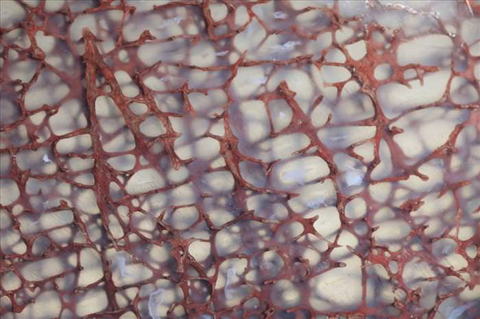 |
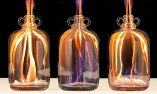
| These dinosaur bones were preserved in a substance called agate, which is a kind of clear silica. Scientists are observing the proteins in an attempt to recreate dinosaur DNA and understand our evolutionary place in relation to theirs. This is a particularly difficult mission because the bones are millions of years old and in may cases are not entirely preserved. Image by: Ted Kinsman |
|
|
|
Nerve in Muscle Section |
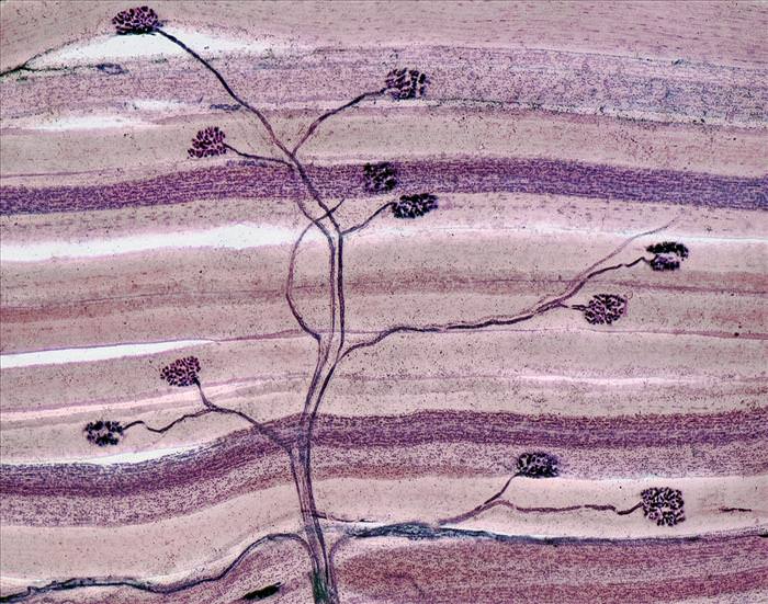 |
| No, this is not a traditional Japanese arboreal painting. This image of nerves traveling through the muscles was taken by sectioning at a magnification of 40x. Nerves are what help us move our muscles, either as a command from the brain, carried down the spinal cord, or as a reaction to elements sensed in the environment. The different points on the nerve force the muscle tissue to move, and are the reasons that we are able to skip, hop and jump through life. Image by: David Ward |
|
|
|
Neuron Receiving Excitatory Contacts |
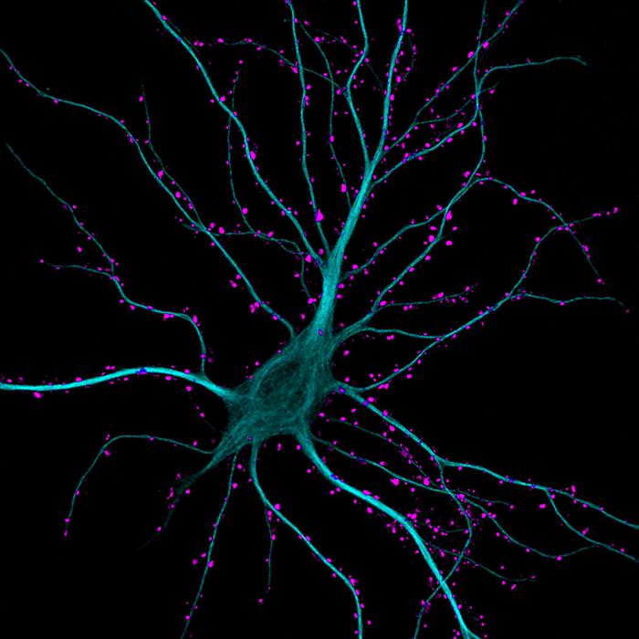 |
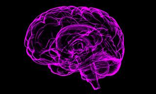
| Neurons have it good; they are always waiting to be excited! When neurons are excited, they are receiving or transmitting information from the brain instantaneously and translating them into physical actions in the muscle tissue. Neurons are really our electrical current, as they carry voltages of information between them. This image does beautiful justice to the electrifying powers of the neuron with its bright fluorescent colors. Image by: Kieran Boyle |
|
|
Submitted by user: Bobby T.